

Kenko Prostate

🔴 < Kenko Prostate

Turning multiparametric prostate MRI into quantitative, explainable clinical insights

What is Kenko Prostate

A next-generation approach to understanding prostate tumour behaviour through complementary MRI biomarkers.

Prostate cancer is the second most frequently diagnosed cancer in men worldwide and the fifth leading cause of cancer death among men. Prostate MRI plays a central role in detection, localisation, and risk assessment, particularly for clinically significant disease however, its interpretation remains complex, time, consuming, and highly dependent on radiologist expertise.

A multiparametric prostate MRI exam generates hundreds of images across sequences such as T2-weighted imaging, diffusion-weighted imaging (DWI), and dynamic contrast-enhanced (DCE) MRI. Radiologists must integrate multiple signal patterns under time pressure, often relying on qualitative frameworks. As imaging demand increases and specialist capacity remains limited, variability and uncertainty continue to create a bottleneck in prostate cancer pathways.

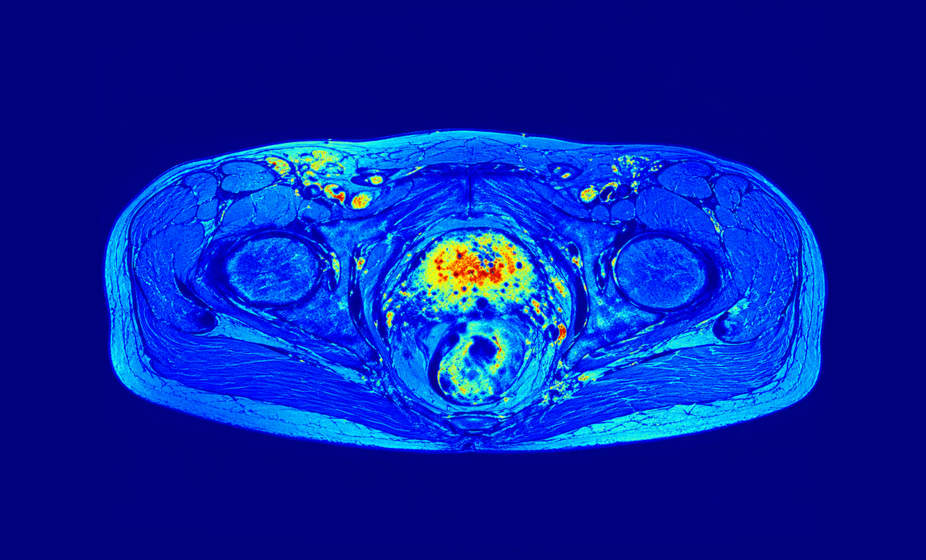
Kenko Prostate addresses this challenge by transforming multiparametric MRI into quantitative, explainable imaging biomarkers that are visual, objective, and clinically interpretable. It provides structured insights into tumour presence, localisation, and biological behaviour, supporting radiologists in reducing reading time, improving consistency, and strengthening confidence in clinically significant cases.

The result is more robust risk stratification, clearer multidisciplinary communication, and more efficient, evidence-based prostate cancer care.

Kenko Prostate

Designed for the
radiologist’s workflow

How Kenko Prostate Works

Kenko Prostate  integrates seamlessly into routine Prostate MRI interpretation, supporting radiologists without changing how exams are acquired or read. After MRI acquisition, the radiologist securely uploads the multiparametric MRI study to Kenko Prostate, where the system processes the full exam and generates quantitative, explainable imaging biomarker maps and structured outputs.

Radiologists review these biomarker maps alongside the original MRI images, enabling clearer lesion characterisation, more consistent interpretation, and stronger evidence for multidisciplinary tumour board discussions. By transforming complex MRI data into visually interpretable, physiology-linked insights, Kenko Prostate helps radiologists move from image overload to quantitative clarity while maintaining full clinical control over interpretation and reporting.

A New Standard in Prostate
MRI Interpretation

From detection to understanding tumour behaviour

Clinical performance

See all

Clinical performance

Narrowing the gap between detection and diagnostic confidence.

Fewer false positives. Fewer missed cancers.
Quantitative imaging biomarkers improve lesion characterisation and diagnostic consistency.

Quantitative evidence, not probability scores.
Physiology-linked biomarkers reveal vascularisation, cellularity, and tumour heterogeneity.

Radiologists

See all

Radiologists

Faster MRI interpretation
Transform numerous MRI images into clear biomarker maps that accelerate reading and reporting.

Objective evidence for complex cases
Quantitative biomarkers reduce inter-reader variability and support confident interpretation.

Stronger tumour board discussions
Visual, explainable biomarker maps provide evidence radiologists can present and defend.

Hospitals and Health Systems

See all

Hospitals and Health Systems

Clinical excellence with operational efficiency
• Reduced unnecessary biopsies and follow-up procedures
• Faster reporting and optimised imaging workflows
• Improved diagnostic consistency across radiologists
• Strategic differentiation in advanced Prostate imaging

Multidisciplinary Oncology Teams

See all

Multidisciplinary Oncology Teams

Built for tumour boards

Kenko Prostate  generates shared quantitative biomarker maps that allow radiologists, oncologists, surgeons, and pathologists to discuss tumour behaviour using the same objective visual evidence.

This supports:

  • Earlier treatment decisions
  • Clearer clinical alignment
  • Reduced uncertainty in complex case

Clinical performance

See all

Clinical performance

Narrowing the gap between detection and diagnostic confidence.

Fewer false positives. Fewer missed cancers.
Quantitative imaging biomarkers improve lesion characterisation and diagnostic consistency.

Quantitative evidence, not probability scores.
Physiology-linked biomarkers reveal vascularisation, cellularity, and tumour heterogeneity.

Radiologists

See all

Radiologists

Faster MRI interpretation
Transform numerous MRI images into clear biomarker maps that accelerate reading and reporting.

Objective evidence for complex cases
Quantitative biomarkers reduce inter-reader variability and support confident interpretation.

Stronger tumour board discussions
Visual, explainable biomarker maps provide evidence radiologists can present and defend.

Hospitals and Health Systems

See all

Hospitals and Health Systems

Clinical excellence with operational efficiency
• Reduced unnecessary biopsies and follow-up procedures
• Faster reporting and optimised imaging workflows
• Improved diagnostic consistency across radiologists
• Strategic differentiation in advanced Prostate imaging

Multidisciplinary Oncology Teams

See all

Multidisciplinary Oncology Teams

Built for tumour boards

Kenko Prostate  generates shared quantitative biomarker maps that allow radiologists, oncologists, surgeons, and pathologists to discuss tumour behaviour using the same objective visual evidence.

This supports:

  • Earlier treatment decisions
  • Clearer clinical alignment
  • Reduced uncertainty in complex case

While most AI tools focus on finding lesions, Kenko Prostate helps clinicians understand tumour behaviour, enabling faster, clearer, and more confident decisions across the cancer diagnostics pathway.

The biomarker logic behind Kenko Devices
Kenko Prostate interprets tumour behaviour using complementary MRI biomarkers derived from perfusion, diffusion, and anatomical context
Kenko Prostate goes beyond detecting suspicious regions in Prostate MRI. The system extracts quantitative imaging biomarkers from multiparametric MRI to reveal how tumours behave biologically. By analysing vascular dynamics, tissue cellularity, and anatomical context together, Kenko Prostate helps radiologists interpret tumour behaviour with greater clarity and confidence.
 

Perfusion biomarkers

Perfusion biomarkers

Understanding vascular behaviour

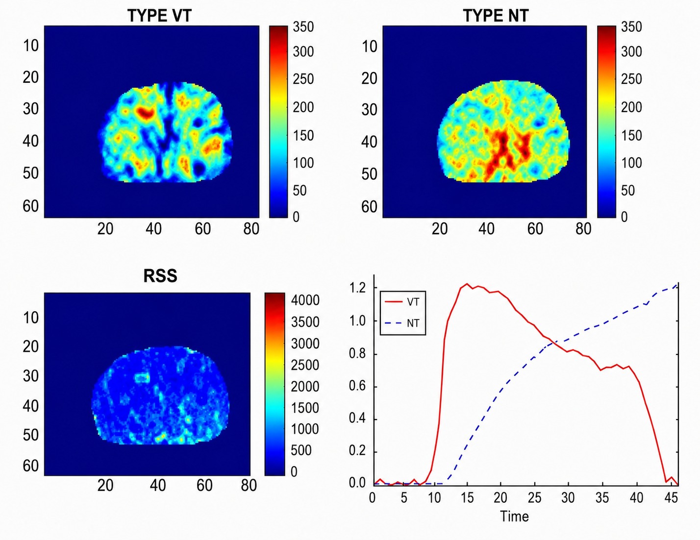
Dynamic contrast-enhanced MRI captures how blood flows through tissue. Kenko analyses both standard and ultrafast contrast dynamics to identify patterns associated with tumour vascularisation and aggressiveness. Early contrast uptake behaviour provides valuable insights into tumour activity and helps distinguish suspicious lesions.
Diffusion biomarkers

Diffusion biomarkers

Understanding tissue cellularity

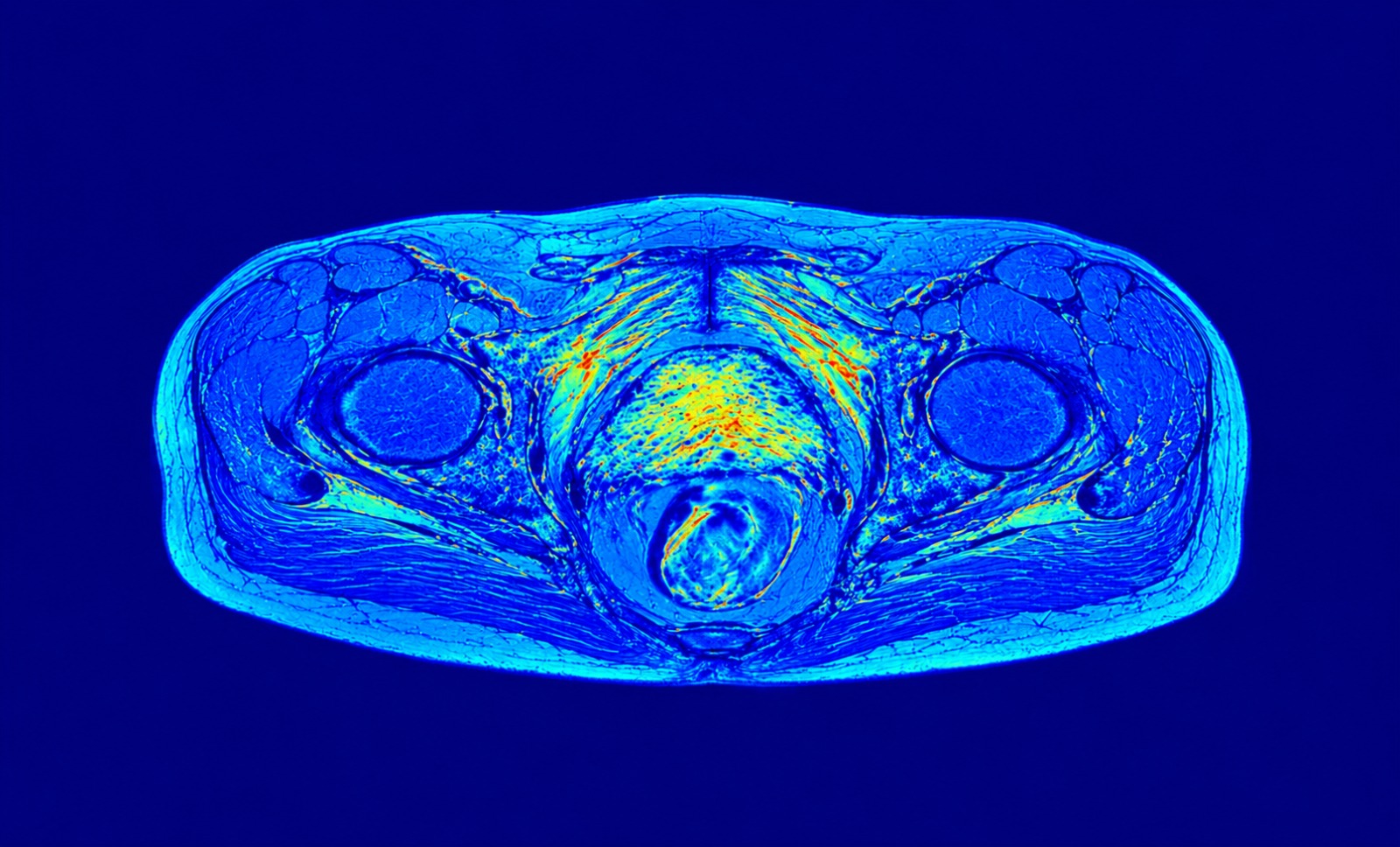
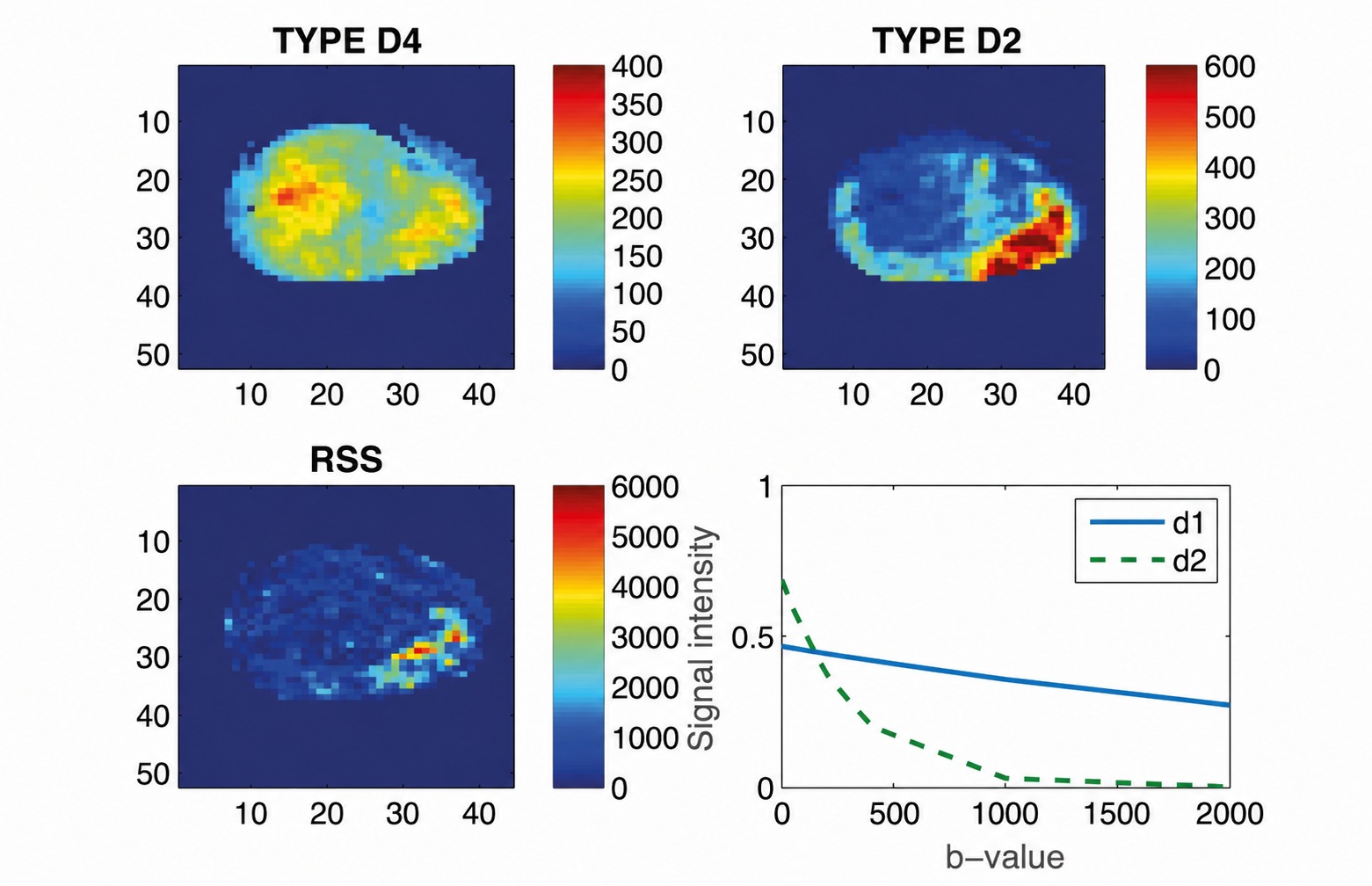
Prostate tissue has an isotropic structure that is analysed through DWI, which is further translated by Kenko Prostate into physiological behaviours and corresponding imaging biomarkers that reflect both tissue cellularity and pseudo perfusion; at the same time reducing noise and improving robustness, which makes Kenko Prostate diffusion information clinically usable for prostate MRI interpretation.

The biomarker logic behind Kenko Devices
Kenko Prostate interprets tumour behaviour using complementary MRI biomarkers derived from perfusion, diffusion, and anatomical context.
Kenko Prostate goes beyond detecting suspicious regions in Prostate MRI. The system extracts quantitative imaging biomarkers from multiparametric MRI to reveal how tumours behave biologically. By analysing vascular dynamics, tissue cellularity, and anatomical context together, Kenko Prostate helps radiologists interpret tumour behaviour with greater clarity and confidence.
 

Perfusion biomarkers

Perfusion biomarkers

Understanding vascular behaviour

Contrast-enhanced MRI captures how blood flows through tissue. Kenko analyses both standard and ultrafast contrast dynamics to identify patterns associated with tumour vascularisation and aggressiveness. Early contrast uptake behaviour provides valuable insights into tumour activity and helps distinguish suspicious lesions.
Diffusion biomarkers

Diffusion biomarkers

Understanding tissue cellularity

Prostate tissue has an anisotropic structure that is not fully captured by conventional diffusion imaging. Kenko uses diffusion tensor imaging (DWI) and advanced modelling to extract stable biomarkers that reflect tissue cellularity and structural organisation. By reducing noise and improving robustness, Kenko makes diffusion information clinically usable for Prostate MRI interpretation.

By combining complementary imaging biomarkers, Kenko Prostate helps radiologists move from detecting lesions to understanding tumour behaviour.

Product Capabilities

What Kenko Prostate can do

Kenko Prostate provides a set of clinically focused functionalities designed to support radiologists in Prostate MRI interpretation, reporting, and multidisciplinary decision-making through the interpretable use of quantitative imaging biomarkers derived from multiparametric MRI. 

Analytical Functionalities

Quantitative Imaging Biomarker Maps

Converts Prostate MRI into pixel-wise quantitative imaging biomarker maps reflecting key tissue properties such as cellularity and vascular behaviour, enabling objective and reproducible assessment beyond qualitative visual MRI reading.

Perfusion Analysis (DCE-MRI)

Performs integrated analysis of dynamic contrast-enhanced sequences, charaterising vascular behavior across temporal phases of contrast uptake and washout, extracting out the structural noise induced by the MR equipment.

Diffusion Analysis (DW-MRI)

Processes diffusion weighted sequences, characterizing the curves in terms of pure diffusion (low exponential decays) and pseudo-perfusion (fast exponential decays), extracting clinically meaningful imaging biomarkers rented to tissue cellularity.

Synchronous Navigation Across Biomarker Maps

Allows radiologists to navigate synchronously across biomarker maps derived from multiple MRI sequences, ensuring consistent spatial correspondence during image review.

Three-Dimensional Functional Visualisation

Provides volumetric visualisation of quantitative biomarker maps to assess tumour extent, spatial distribution and heterogeneity.

Selective Maximum Intensity Projection (Selective MIP)

Generates maximum intensity projection views of biomarker maps and allows radiologists to define the slice range used for projection.

Quantitative Lesion and Tissue Characterisation

Supports quantitative characterisation of Prostate lesions and surrounding tissue based on imaging biomarker maps derived from multiparametric MRI.

Together, these capabilities transform complex multiparametric Prostate MRI into quantitative, interpretable evidence that supports clearer interpretation, consistent reporting and confident multidisciplinary decision-making.

Check your email for your login credentials to
access Kenko Breast AI+.

Your login details have been sent to the email address linked to your authorised account. Please
follow the instructions included in that email to
access the platform securely.

Thank you.